MSACL 2023 Abstract
Self-Classified Topic Area(s): Imaging > Metabolomics
|
|
 Probing 3D Mammalian Co-culture Models of Ovarian Cancer Metastasis for Metabolic Crosstalk Using Imaging Mass Spectrometry Probing 3D Mammalian Co-culture Models of Ovarian Cancer Metastasis for Metabolic Crosstalk Using Imaging Mass Spectrometry
Hannah Lusk (1), Monica Haughan (2), Tova Bergsten (2), Joanna E Burdette (2), Laura M Sanchez (1)
(1) Department of Chemistry and Biochemistry, UC Santa Cruz, Santa Cruz, CA, 95064
(2) Department of Pharmaceutical Sciences, University of Illinois at Chicago, Chicago, IL 60612
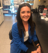 | Hannah Lusk, PhD (Presenter) 
University of California San Francisco | Presenter Bio: I earned my B.Sc. and M.Sc. degrees in Biochemistry from Kansas State University in 2019, where I conducted research in Dr. Ruth Welti’s lab on genes involved in lipid metabolism in Arabidopsis thaliana. In 2024, I completed my Ph.D. in Chemistry at the University of California, Santa Cruz under Dr. Laura Sanchez, where my dissertation focused on chemical communication in mammalian systems using MS-based metabolomics. I am currently a Clinical Chemistry Fellow at the University of California, San Francisco, working with Drs. Alan Wu and Kara Lynch to develop and validate mass spectrometry–based assays for toxicology and therapeutic drug monitoring, with a focus on anti-tuberculosis drug monitoring and emerging drugs of abuse.
| Grant/Research Support |
NIH |
|
|
|
|
|
|
Abstract INTRODUCTION:
Ovarian cancer is the most lethal gynecologic malignancy, and high-grade serous ovarian cancer (HGSOC) is responsible for 70-80% of ovarian cancer deaths. HGSOC originates in the fallopian tube; it then metastasizes, first to the ovary during primary metastasis, then to the omentum during secondary metastasis. We hypothesize this pattern of metastasis is governed by the exchange of small molecules between ovarian cancer cells and the tissues they metastasize to. To investigate this, we developed an imaging mass spectrometry protocol for analyzing 3D co-cultures of mammalian cells and healthy murine tissue explants. We previously used this protocol with ovarian tissue to probe chemical signaling in primary metastasis, and have recently adapted it for use with omental tissue to investigate secondary metastasis.
OBJECTIVES:
The primary objectives of this study are to identify small molecules exchanged between ovarian cancer cells and omental tissues, and to characterize their role in metastasis.
METHODS:
Healthy murine omental tissues were co-cultured with 1) fallopian tube epithelial cells (FTEs) that have an shRNA PTEN mutation that results in tumor formation in the murine model, 2) healthy FTEs, and 3) murine ovarian surface epithelial cells in an agarose-based 3D mammalian cell culture medium. We have also been exploring the use of omental organoids alongside omental explants to validate some of the findings at the genetic level. Matrix-assisted laser desorption/ionization-quadrupole time-of-flight imaging mass spectrometry (MALDI-Q-TOF IMS) was used to compare the spatial distributions of metabolites in these co-cultures to identify small molecule metabolites specific to the omentum/tumorigenic FTE condition. Metabolites are orthogonally validated using ultra-high performance liquid chromatography-trapped ion mobility-tandem mass spectrometry (UPLC-TIMS-MS/MS) on a Bruker timsTOF fleX mass spectrometer. Putative ID’s are validated by matching fragmentation patterns and retention times to commercial standards or databases.
RESULTS:
To adapt our existing protocol for use with omental tissue, a number of alterations were required. MALDI-based IMS requires a homogeneously flat sample; unfortunately, due to its fatty nature, omental tissue did not dry flat but rather crystallized when desiccated using heat. To circumvent this issue, the protocol has been adapted by placing tissue in the corner of the agarose plug and removing it prior to desiccation. Additionally, agar and agarose-based samples can be difficult to consistently prepare as they are susceptible to humidity and temperature fluctuations, which can lead to inconsistent drying and wrinkling on the sample surface. Towards facilitating the drying process, we recently reported the construction of a home-built spinning apparatus to rotate samples as they dry, making the process of using agar and agarose-based samples more robust and less user error-prone. Our data demonstrate that robotic spinning helps samples dry more evenly, and accelerates the drying process. This new protocol has facilitated the identification of more than 50 signals specific to cancer cell/omentum co-culture conditions in both positive and negative mode. Our preliminary data point to alterations in methionine and proline metabolism, as well as the activation of lipolysis and β-oxidation in cancer cell/omentum co-culture conditions. We are currently working to validate these signals and determine their origin (cancer cells or omentum) using a Bruker timsTOF fleX mass spectrometer. Future work will focus on characterizing the functional role of specific metabolites in the context of ovarian cancer metastasis.
CONCLUSIONS:
We have a new protocol for imaging the secondary metastasis of HGSOC in vitro using whole omental tissue; it has been used to identify over 50 signals specific to cancer cell/omentum co-culture conditions. Putatively identified metabolites are currently being validated using a Bruker timsTOF fleX mass spectrometer. The identification of small molecules exchanged at this interface could unearth key pathways whose role in HGSOC is poorly understood.
|
|
| |
 = Translation stage. (39.50%, 2023)
 = Clinically available. (36.13%, 2023)
