MSACL 2025 AbstractKeynote Presentation
Self-Classified Topic Area(s): Spatialomics > Spatialomics : Pathology and Biomarkers > Emerging Technologies
|
|
 Challenging Precision Medicine and Surgery in Oncology with MS Imaging-guided Spatial Omics Combined with AI Challenging Precision Medicine and Surgery in Oncology with MS Imaging-guided Spatial Omics Combined with AI
Isabelle Fournier (1,2)
(1) PRISM Inserm U1192 University of Lille, 59000 Lille, France
(2) Institut Universitaire de France, Paris, France
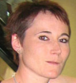 | Isabelle Fournier, PhD (Presenter)
Laboratoire PRISM - Université de Lille | Presenter Bio: Distinguished Professor I. Fournier (IF, PRISM, U1192, Lille) is senior member of the University Institute (IUF) with a chair in Clinical Mass Spectrometry. She is currently co-director of PRISM lab INSERM U1192. Prof. Fournier is a specialist of Mass Spectrometry applied to clinics and proteomics. She was pioneer a novel technology in Europe of molecular imaging namely MALDI Mass Spectrometry Imaging in 2002 (Fournier et al., Neuroend. Letters 2003) and in 2007 demonstrated that MALDI MS Imaging could be perform from archived Formalin fixed and Paraffin Embedded hospital tissue samples (Lemaire et al., J. Proteome Res 2007). In 2005, she developed the multiplex targeted imaging, Tag-mass technology (Lemaire J. Proteome Res. 2007; Gagnon et al., Prog Histochem Cytochem 2012). In 2013, she introduced the concept of the spatially-resolved tissue proteomic and applied this technology on different cancers (Longuespee et al., Cancer Metastasis Rev 2012; Quanico et al Chem Com, 2015, Simeone et al., Semin Cancer Biol., 2018). Since 2011, Pr. I. Fournier with Pr. X. Roucou have evidenced the Ghost proteome (Samandi et al., Elife 2017, Delcourt et al., EBioMedicine et al., 2017, Brunet et al., Nucleic. Acid. Res., 2019). In 2014, she performed the development of a novel technology based on mass spectrometry and allowing to realize in vivo real time tissue molecular imaging and for guiding surgery, with an instrument named SpiderMass (Saudemont et al., Cancer cells, 2018; Ogrinc et al., Nature Protocols, 2019).Over the past 5 years, she also worked on the development of in vivo MS for guided surgery and intraoperative analysis. She confounded the Imabiotech Company which provides services in MALDI MSI and recently CELEOS that will commercialize the SpiderMass technology. She was recently distinguished by the international distinguish award from MSACL and was awarded a senior position at Institut Universitaire de France in 2019.
No relevant financial relationship(s) to disclose.
|
|
|
|
|
|
Abstract Large-scale MS-based omics has revolutionized the field of metabolomics and proteomics. However, conventional strategies lack the spatial dimension, crucial to better understand physiological and pathophysiological mechanisms by accessing the cell microenvironment. On the other hand, MS imaging (MSI) brings the spatial distribution of molecules down to the single cell, but fails to provide large-scale identification. Therefore, bridging MSI and large-scale omics to perform MSI-guided spatial omics opens new avenues for elucidating biological mechanisms and their clinical translation. In this regard, we have developed various approaches for spatial omics from histological tissue sections, including microextraction-based for both shotgun and top-down proteomics, or laser ablation-based using backside irradiation. These approaches have been applied to clinical questions such as stratification of glioblastoma patients, discovery of diagnostic and prognostic markers, and understanding the early onset of ovarian cancer. To increase the depth of spatial information, laser ablation can be automated, allowing an entire section of tissue to be sampled according to a defined grid of ablation points, which can then be processed through a pipeline suitable for small amounts of material to obtain protein identification and relative quantification. Label-free quantification values are then used to obtain images of thousands of identified proteins. Such a strategy is being developed to access down to 50 µm spatial resolution and provide complete proteome information at the scale of clonal heterogeneity for oncology. To bring these approaches closer to the patient's bedside and to make them useful in clinical routine, we have also developed the concept of dried proteomics. This concept involved machine learning to train a model for a specific type of clinical sample to associate the image of lipids with the protein pathways. Thanks to this new concept, it is then possible to predict the patient's overall survival and evolution, or to test the adequacy of the treatment used when it is performed on patient organoids.
Finally, to open MS to more applications, limit sample preparation, and advance in vivo measurements, we have developed a novel ambient ionization mass spectrometry (AIMS) technology. The SpiderMass uses the body's water molecules to achieve desorption/ionization with minimal invasiveness. By connecting the laser probe to a robotic arm, SpiderMass can be operated in MSI mode to generate topographic MS images, predict immune cell infiltration, and generate immunoscores and bacterioscores that can be obtained directly at the time of initial surgery. |
|
| |
 = Discovery stage. (53.14%, 2025)
 = Clinically available. (24.53%, 2025)
