MSACL 2025 Abstract
Self-Classified Topic Area(s): Spatialomics > Spatialomics : Automation and AI > none
|
|
 Advancing Spatial Pathomics with MALDI MSI from Imaging to Insights Advancing Spatial Pathomics with MALDI MSI from Imaging to Insights
Jader Giraldo-Guzmán (1), Cody Weimholt (2), Lewis J. Thomas (3), Sheng-Kwei Song (1), Peggi M. Angel (4), Richard R. Drake (4), Joseph E. Ippolito (1,5), José Marcio Luna (1)
(1) Mallinckrodt Institute of Radiology, Washington University in St. Louis, USA, (2) Department of Pathology and Immunology, Washington University in St. Louis, USA, (3) Department of Surgery, Washington University in St. Louis, USA, (4) Department of Pharmacology and Immunology, Medical University of South Carolina, USA, (5) Department of Biochemistry and Molecular Biophysics, Washington University in St. Louis, USA
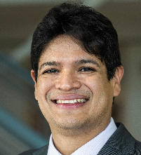 | Jose Marcio Luna, MS, PhD (Presenter)
Washington University in St. Louis | Presenter Bio: Dr. José Marcio Luna is an Assistant Professor at the Mallinckrodt Institute of Radiology at Washington University in St. Louis. He leads a research program focused on advancing cancer diagnosis and prognostication through innovative data integration methods, bringing expertise in cancer research, machine learning, data sciences, and computational medical imaging. Dr. Luna earned his M.S. and Ph.D. (with a Ph.D. minor in Applied Mathematics) in Engineering from the University of New Mexico in 2010 and 2014, respectively, where he focused on modeling intrinsically random nonlinear systems. In 2021, he completed postdoctoral training in Radiology at the University of Pennsylvania, developing AI-based methods to predict treatment effectiveness in non-small cell lung cancer (NSCLC) and contributing to interpretable machine learning methods for medical applications. Dr. Luna received the 2021 Marlene Shlomchik Fellowship and an Emerson Collective grant during his postdoc. With 36 peer-reviewed publications, including 12 in prestigious journals such as PNAS and Nature Scientific Reports, Dr. Luna is now leading a research initiative funded by an NCI Transition Career Development Award. His work focuses on identifying prognostic biomarkers from radiological and histopathological imaging to enhance treatment guidance and prognostication for various cancers.
No relevant financial relationship(s) to disclose.
|
|
|
|
|
|
Abstract INTRODUCTION:
Prostate cancer (PCa) progression is significantly influenced by interactions between extracellular matrix (ECM) proteins within the stroma and malignant epithelial cells. Despite their critical role in tumor biology, these interactions are not currently considered in the standard clinical evaluation of PCa. Previously, our team identified ECM glycans associated with PCa using matrix-assisted laser desorption/ionization (MALDI) imaging, providing preliminary evidence that MALDI can guide pathomic analysis in hematoxylin and eosin stain (H&E) images. However, manual registration between MALDI and H&E images limited its clinical applicability. Here, we present a fully automated, robust co-registration and segmentation pipeline that enables quantitative pathomic analysis of MALDI-guided regions in H&E images and extends to multi-modality integration with ex vivo T2-weighted Magnetic Resonance Imaging (T2W MRI). Unlike MALDI, which provides spatial maps of molecular features, T2W MRI captures anatomical contrasts based on tissue water content, making their co-registration a non-trivial computational challenge.
OBJECTIVES:
Our primary goal is to automate image co-registration and segmentation, allowing for precise identification of pathomic textural features guided by MALDI-derived glycan expression in H&E images. Additionally, we aim to extend this automation to 54 T2W MRI sectors, demonstrating the feasibility of integrating histopathology, MALDI, and ex vivo MRI for comprehensive tissue characterization. We further explore the robustness of our pipeline by quantifying co-registration quality across these distinct imaging modalities.
METHODS:
We analyzed a cohort of PCa patients (n=10) who had undergone radical prostatectomy with Gleason grade groups GG3–GG5. For each patient, H&E images, five MALDI images representing distinct N-glycan species (1419, 2539, 2686, 1663, and 1809 m/z), and 54 ex vivo T2W MRI sectors were obtained from the cohort, with varying numbers of sectors contributed by individual patients. Both the MALDI and T2W MRI datasets originated from the same 10 patients, allowing for direct multi-modality co-registration and comparison. Co-registration was fully automated using feature-based deformable registration methods with mutual information metrics, minimizing parameter tuning and eliminating the requirement for data-driven training. For H&E, segmentation was performed using a computational imaging framework involving rolling ball, erosion, dilation, Otsu’s thresholding, and connected component analysis, resulting in six regions of interest: five MALDI-derived N-glycan regions and one pathologist annotation. Pathomic features (n=70) were extracted from the H&E images within the six co-registered regions of interest using PyRadiomics. Wilcoxon signed-rank tests were performed to compare textural differences across these specific regions. Furthermore, we applied the same automated co-registration method, to align MRI T2W and H&E images. Quantitative evaluation of co-registration quality was performed using Dice similarity coefficients, assessing the spatial overlap between MALDI, H&E and T2W MRI of whole tissue sections.
RESULTS:
The median Dice scores for each MALDI-derived glycan against pathology annotations were: 1419 m/z (0.48 [0.39–0.65]), 2539 m/z (0.64 [0.44–0.76]), 1663 m/z (0.27 [0.15–0.37]), 1809 m/z (0.38 [0.14–0.42]), and 2686 m/z (0.29 [0.09–0.35]). This indicates that glycans 1419 and 2539 m/z have the greatest overlap with tumors, while glycans 1663, 2686, and 1809 m/z predominantly overlap with non-tumor regions. Out of the 70 pathomic textural features, 55 were significantly different (p < 0.05) between the masks for 1419 and 1663 m/z, 48 between 1419 and 1809 m/z, 47 between 2539 and 1663 m/z, and 50 between 2539 and 1809 m/z. In contrast, only 10 features were significantly different between 1419 and 2539 m/z, and 4 features between 1663 and 1809 m/z. These pathomic results are consistent with the categorization of tumor and non-tumor glycans indicated by the Dice scores. In addition, automated co-registration significantly improved overlap quality for whole tissue sections, with a median relative Dice score increase of 57.88% [41.79%–88.83%] when co-registering the entire MALDI tissue region with the whole tissue H&E slide. The process was further extended to 54 T2W MRI sectors, where the entire tissue from the MRI was co-registered with the whole tissue on the H&E slide, achieving a median relative Dice score increase of 47.07% [28.09%–66.63%]. This demonstrates the robustness of our pipeline across different imaging modalities and scales.
CONCLUSIONS:
We demonstrate the feasibility of a fully automated, robust co-registration and segmentation pipeline for MALDI-guided pathomic analysis of H&E images in PCa. Our results provide evidence that MALDI imaging not only identifies regions of tumor involvement but also enables precise pathomic quantification without manual intervention. Moreover, the extension of this approach to T2W MRI, despite the fundamental differences in image contrast, showcases the adaptability of our pipeline for multi-modality integration, potentially streamlining clinical workflows for prostate cancer diagnostics. Future work will focus on clinical translation, assessing these biomarkers in larger cohorts, and validating the pipeline in clinical laboratory settings.
|
|
| |
 = Translation stage. (22.33%, 2025)
 = Clinically available. (24.53%, 2025)
