MSACL 2026 Abstract
Self-Classified Topic Area(s): Proteomics > Tox / TDM / Endocrine
|
|
 QconCAT Proteomics of Human Adrenals for Absolute Quantitation of Cytochrome P450 Enzymes QconCAT Proteomics of Human Adrenals for Absolute Quantitation of Cytochrome P450 Enzymes
Heather A. Hartmann (1), Kevin D. McCarty (1), Sarah M. Glass (1), W. Hayes McDonald (2), Richard J. Auchus (3), and F. Peter Guengerich (1)
(1) Department of Biochemistry, Vanderbilt University School of Medicine, Nashville, Tennessee, USA
(2) Mass Spectrometry Research Center, Vanderbilt University School of Medicine, Nashville, Tennessee, USA
(3) Division of Metabolism, Endocrinology, and Diabetes, University of Michigan, Ann Arbor, Michigan, USA
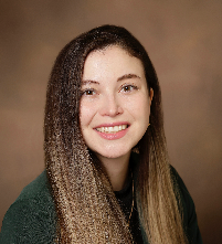 | Heather Hartmann, Ph.D. (Presenter) 
Vanderbilt University | Presenter Bio: I am in my second year of my post doc studies in the lab of Dr. F. Peter Guengerich at Vanderbilt University in Nashville, TN, where my main research focuses on inhibiting the interaction of P450 17A1 and cytochrome b5 in prostate cancer. I completed my PhD at Vanderbilt University in 2025 in the laboratories of Dr. Vivian Weiss and Dr. Ethan Lee, where the main research focus is understanding how Tenascin-C interacts with the Wnt signalosome. I received a BA in Spanish and Chemistry at the College of Wooster in 2021, where my research focused on mass spectrometry methods for environmental contaminants. My research interests during my post doc are expansive in the field of cancer biology, P450s, and mass spectrometry to understand more about cancer and drug metabolism.
No relevant financial relationship(s) to disclose.
|
|
|
|
|
|
Abstract INTRODUCTION:
The human adrenal glands are the primary source of the essential steroid hormones cortisol and aldosterone, as well as a secondary source of androgens and estrogens. Critical reactions are catalyzed by cytochrome P450 enzymes (P450s, CYPs) dispersed throughout three regions of the adrenal cortex: the zona glomerulosa, zona fasciculata, and zona reticularis. Each zone corresponds to differences in steroidogenesis: the zona glomerulosa produces aldosterone from cholesterol via P450s 11A1, 21A2, & 11B2; zona fasciculata produces cortisol via P450s 11A1, 17A1, 21A2, and 11B1; and zona reticularis produces androgen precursors via P450s 11A1 and 17A1. The cytochrome P450s can also be classified by subcellular location: mitochondria (P450s 11A1, 11B1, 11B2, which utilize redox partners adrenodoxin and adrenodoxin reductase) or microsomal (P450s 17A1, 21A2, which utilize NADPH-cytochrome P450 reductase and cytochrome b5). While immunohistochemical studies have identified the zonal localization of several of these P450s and their redox partners, further work is necessary to quantify the protein levels. Understanding physiological P450 and redox partner concentrations can help refine in vitro assays by informing stoichiometry of redox partners and P450s and reveal alterations of P450 levels in adrenal diseases.
METHODS:
A QconCAT containing 2 peptides for each protein of interest was expressed in E. coli in minimal media with 13C6-lysine and 13C6-arginine and then purified using metal affinity chromatography. Human adrenal tissue from 14 donors (7 males and 7 females) were pooled, homogenized, and solubilized before QconCAT peptide was added. The tissue homogenate and QconCAT sample were then digested using trypsin, and two technical replicates of our adrenal samples were analyzed by LC-MS/MS. We performed quantitation of P450s and redox partners via comparison of peak areas for peptides in homogenized tissue digests with those of heavy peptide internal standards.
RESULTS:
The P450 redox partner protein adrenodoxin (Adx) was the most abundant of our targeted proteins with an amount of 20±2 pmol/mg of total protein. The most abundant microsomal P450 was P450 17A1 (9±1 pmol/mg). The quantification for the remaining proteins were as follows: P450 11A1 (7±1 pmol/mg), P450 11B1 (6±1 pmol/mg), P450 21A2 (3±1 pmol/mg), adrenodoxin reductase (5±1 pmol/mg), cytochrome P450 reductase (4±4 pmol/mg), cytochrome b5 (0.9±0.3 pmol/mg), and P450 11B2 (0.6±0.1 pmol/mg).
DISCUSSION:
We determined the total, net abundance of P450s and redox partners in adrenal homogenates. P450 17A1 was the most abundant P450 while P450 11B2 was the least abundant protein. Our preliminary results suggest that physiological values for in vitro assays might be: Adx:P450 ratios around 33:1 for 11B2 or 3:1 for 11B1 and 11A1. Absolute and relative quantification of proteins in each zone, however, is necessary to draw firm conclusions. An ongoing approach is to determine the abundance of each P450 and redox partner within each zone of the adrenal using laser capture microdissection and imaging mass spectrometry. Determining the abundance of each P450 and their redox partner proteins will help to further determine stoichiometries and differences in their abundances in adrenal diseases. |
|
| |
 = Translation stage. (23.38%, 2026)
 = Clinically available. (19.40%, 2026)
