A Focused Agenda for Pathologists Interested in Spatial Mass Spectrometry
Tuesday - Thursday, March 19-21
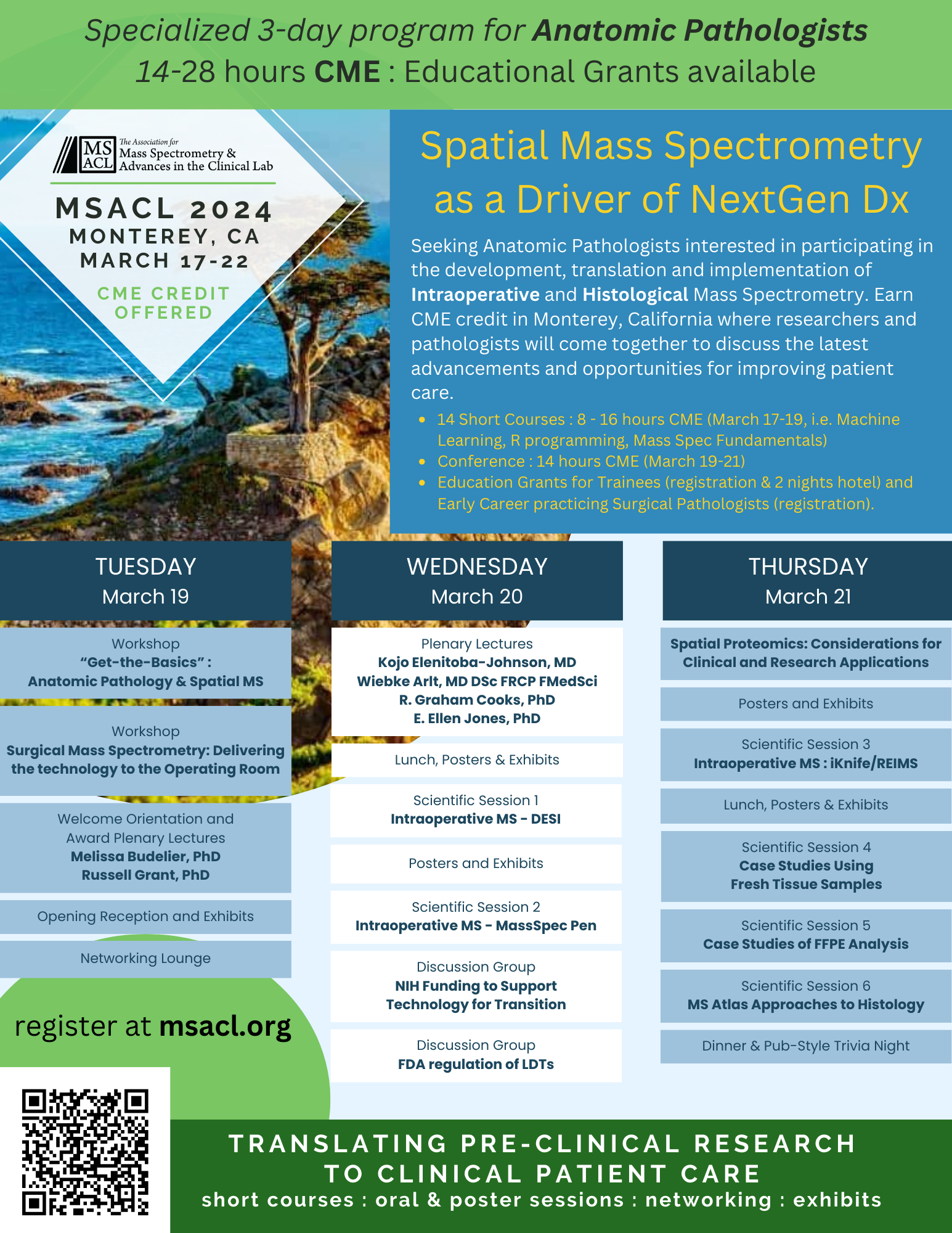
Seeking Anatomic Pathologists interested in participating in the development, translation and implementation of Intraoperative and Histological Mass Spectrometry.
Earn CME credit in Monterey, California where researchers and pathologists will come together to discuss the latest advancements and opportunities for improving patient care.
MSACL 2024 and members of the Mass Spectrometry Imaging community are building a specialized 3-day program, March 19-21, on Spatial Mass Spectrometry as a Driver of NextGen
Dx (see below).
Educational grants are available for Trainees (registration and 4 nights hotel) and practicing early career Pathologists (registration). Apply for educational grants.
Exhibit booths and industry workshops are available.
Please share with your local Anatomic Pathologists and MSI friends!
Can't make it to Monterey? Mark September 21-26, 2025 on your calendar for the next edition to be held in Montreal, Canada.
click image to view/copy full-size image
click image to view/copy full-size image
Time
Sessions
Tuesday 945
1045
Workshop : Concepts in Histology and Histopathologic Diagnosis for Researchers
Location: De Anza 1 (Portola Hotel > Ground Floor)
Albert Tsai, MD, PhD
Stanford
Dr. Tsai received his undergraduate training at the University of California, Los Angeles (B.S., Biochemistry, summa cum laude), followed by combined medical and graduate training at the University of Southern California (M.D., Ph.D., Biochemistry). He completed anatomic and clinical pathology (AP/CP) residency and hematopathology fellowship at Stanford University, receiving board certification in AP/CP and hematopathology. As an instructor, he performed clinical diagnostic duties on the hematopathology service while doing postdoctoral training in the laboratory of Dr. Sean Bendall, with funding from the Damon Runyon Cancer Research Foundation.
As a physician and hematopathologist, he seeks to mechanistically dissect myelodysplastic syndromes (MDS) using highly-multiplexed immunophenotyping — mass cytometry / cytometry by time-of-flight (CyTOF) and multiplexed ion beam imaging (MIBI). MDS is an especially complex and heterogeneous disease of abnormal blood cell development with increasing prevalence and few treatments. By combining practical experience clinically diagnosing MDS, next generation single cell proteomic approaches, fundamental discoveries in the biology of MDS, and knowledge of clinical laboratory testing, we hope to develop new clinical diagnostics for personalizing MDS therapies and therapeutic monitoring.
His clinical diagnostic duties are on the hematopathology service, primarily in the diagnosis of MDS, leukemias, lymphomas, and other hematopoietic diseases from blood, bone marrow, and tissue samples.
How pathologists diagnose human tissue samples differs markedly from how many researchers study them. Research approaches often seek to reduce two-dimensional tissue imaging to single cell data, i.e. cytometry-on-a-slide. However, these methods are complicated by technical aspects of tissue processing, and they may miss the overarching histologic patterns which underlie pathologic diagnosis. Thus, early engagement with pathologists is useful not only for obtaining tissue samples, but also for understanding structure-function relationships and relevance to disease. Furthermore, pathologists regularly integrate multiple data modalities to classify diseases, e.g. histology with antibody-based protein detection and DNA sequencing. Their understanding of the utilities of each modality within the larger diagnostic context is helpful for identifying potential roles for new technologies.
Tuesday 1100
1200
Get-the-Basics : MALDI Mass Spectrometry Imaging – A New Method in Pathology?
Location: De Anza 1 (Portola Hotel > Ground Floor)
Kristina Schwamborn, MD, PhD
Technical University of Munich
Matrix assisted laser desorption ionization (MALDI) mass spectrometry imaging (MSI) combines the excellence in molecular characterization of mass spectrometry with microscopic imaging capabilities of stained tissue samples, enabling the precise location of different analyte classes (e.g., proteins, peptides, lipids, glycans) directly within intact tissue. In particular in the field of pathology, that can aid in tumor diagnosis, tumor subtyping, biomarker identification, prognostic prediction, and characterization of tumor margins during tumor resection procedures. Since MALDI MSI goes far beyond microscopy, it is ideal for these endeavors. It can generate molecular maps of tissue sections that can elucidate the underlying biochemistry or provide information on how therapeutics or toxins influence the function or misfunction of an organ. Thus, it has the potential to overcome limitations of other approaches in the identification and routine diagnostic measurement of new marker molecules/profiles.
Different applications in the field of pathology/ oncology will be presented that highlight possible applications of MALDI MSI in Pathology. Combining MALDI MSI, histology, and statistical analysis allows for reliable and fast subtyping in a number of different tumor types while also conserving material that could be used for additional testing.
Location: San Carlos 4 (Marriott > Mezzanine | Stairs from Lobby or SkyBridge from Conference Ctr)
Tuesday 1400
1600
Workshop : Surgical Mass Spectrometry – Delivering the Technology to the Operating Room
Location: De Anza 1 (Portola Hotel > Ground Floor)
Zoltan Takats, PhD
Imperial College
Professor Takats has obtained his PhD from Eötvös Loránd University, Budapest, Hungary. He has worked as a post-doctoral research associate at Purdue University, Indiana, USA. After returning to Hungary, he served as Director of Cell Screen Research Centre and also as Head of Newborn Screening and Metabolic Diagnostic Laboratory at Semmelweis University, Budapest.
Professor Takats was awarded the Starting Grant by the European Research Council in 2008 and he subsequently, became a Junior Research Group Leader at Justus Liebig University, Gießen, Germany. He moved to the United Kingdom in 2012 and currently works as a Professor of Analytical Chemistry at Imperial College London.
Professor Takats has pursued pioneering research in mass spectrometry and he is one of the founders of the field of ‘Ambient Mass Spectrometry’. He is the primary inventor of six mass spectrometric ionization techniques and author of 78 peer reviewed publications. He was the recipient of the prestigious Mattauch-Herzog Award of the German Mass Spectrometry Society and the Hungarian Star Award for Outstanding Innovators. He is the founder of Prosolia Inc, Medimass Ltd and Massprom Ltd, all companies pursuing analytical and medical device development.
Lauren Ford, BSc (Hons), PhD
Imperial College London
I am an early career researcher and have a background in materials chemistry, having studied for a PhD between the School of Chemistry and the School of Design at the University of Leeds I have experience in polymer technology, physical adsorption theory and purification. I am interested in using these skills to aid detection of disease using mass spectrometry detection. Since joining Imperial in 2019 I have been working as a post-doctoral research associate in the department of Surgery and Cancer, working on the iEndoscope project. This project utilised ambient ionisation mass spectrometry and allowed me to gain critical experience of ambient MS for early cancer detection.
Objectives
To identify methods to deliver mass spectrometry guided surgery to become routinely used in the clinical interventional world. The workshop will focus on the
interpretation of clinical information/data and how this should be fed back to healthcare professionals
and ultimately patients. The workshop will highlight the limitations of current technologies and
developments enabling clinical adoption.
Summary
The need for in-situ, real time tissue identification has been dramatically increasing with the
development and deployment of robotic and other high-precision surgical approaches. While surgical
mass spectrometry techniques have been continuously developed, published, and demonstrated in
human surgical theatres, none of these approaches have reached regulatory approval and routine
application in surgery. We will use this interactive setting to discuss overcoming current roadblocks to
delivering technology for patients and healthcare professionals. The workshop will give an overview of
the current mass spectrometry technology developed and the strengths and weaknesses in each
approach. This will be followed by discussing the embedding of the approach both into existing oncology
and clinical diagnostic systems. Using Mass Spectrometry in surgery will change how interventional
cancer care is delivered, hence it is important to ensure data tools are developed which can be relied on.
Delivery of the obtained clinical information in the operating theatre is also important to explore. As part
of this workshop, we will discuss data visualisation strategies such as in the virtual reality space,
delivery of feedback to the clinical healthcare professionals and tools developed to advance usability,
such as navigation. Data interpretation in the wider context of clinical oncology will also be explored.
Syllabus/Topics
• Surgical mass spectrometry methods – strengths, weaknesses, applications and future perspectives
• Embedding of technology into healthcare systems. How to deliver clinical information, data interpretation, navigation guidance and feedback.
• Regulatory and health economics aspects.
Wednesday 900
950
Plenary : Proteogenomics as a driver for discovery of novel mechanisms and therapeutic targets in lymphoma pathogenesis
Dr. Elenitoba-Johnson is the inaugural Chair of the Department of Pathology and Laboratory Medicine, and Member of the Human Oncology & Pathogenesis Program (HOPP) at Memorial Sloan Kettering Cancer Center. Prior to this appointment, he was the Director of the Center of Personalized Diagnostics, and the inaugural Peter C. Nowell, M.D., Endowed Professor, in the Department of Pathology and Laboratory Medicine at Penn Medicine, Perelman School of Medicine, at the University of Pennsylvania from 2015 to August 2022.
He is a recognized pioneer in lymphoma proteomics, and a top leader in precision and integrated diagnostics. His work is notable for the identification and mechanistic elucidation of targetable genetic alterations underlying the pathogenesis of specific lymphoma subtypes. Dr. Elenitoba-Johnson has contributed to over 180 peer-reviewed manuscripts, numerous chapters and text books. His research is supported by 3 RO1 awards from the National Institutes of Health.
Dr. Elenitoba-Johnson is an elected member of the National Academy of Medicine, the American Society for Clinical Investigation and the International Lymphoma Study Group. (2017) and has been recognized with numerous professional honors and awards, notably the Ramzi Cotran Young Investigator Award from the United States and Canadian Academy of Pathology, the Outstanding Investigator (Former Warner-Lambert-Parke Davis) Award from the American Society for Investigative Pathology and the William Gerald Award from Memorial Sloan Kettering Cancer Center.
Declaration of Competing Interests
Dr. Elenitoba-Johnson declares no conflicts of interest.
Wednesday 1000
1050
Plenary : Steroid Metabolomics for Diagnostic and Prognostic Biomarker Development
Medical Research Council Laboratory of Medical Sciences
Wiebke Arlt is the Director of the Medical Research Council Laboratory of Medical Sciences (LMS) and Professor of Transdisciplinary Medicine at Imperial College London; she also serves as Honorary Consultant Endocrinologist at Imperial College Healthcare Trust, with a clinical focus on adrenal and reproductive endocrinology.
At the LMS, she leads a multi-disciplinary research group comprising biochemists, clinician scientists and computational biologists, investigating the role of steroids in health and disease, with a focus on the link between steroids and metabolism. Her group uses steroid mass spectrometry in combination with in vitro, ex vivo and in vivo phenotyping in humans as a discovery tool and for the development of biomarkers utilised for diagnostic and prognostic test purposes.
Wiebke has published over 250 original research articles and is a sought-after lecturer. Her scientific work has attracted major international prizes, most recently the 2021 Keith Harrison Memorial Lecture of the Endocrine Society of Australia, the 2019 Outstanding Clinical Investigator Award of the Endocrine Society USA, the 2017 Berthold Medal of the German Endocrine Society, and the 2016 Clinical Endocrinology Trust Medal of the European Society of Endocrinology. She was elected Fellow of the UK Academy of Medical Sciences in 2010 and currently serves on the Academy’s Council.
I will discuss steroid metabolomics, the combination of mass spectrometry-based steroid profiling with machine learning-based steroid data analysis. I will provide two clinically relevant examples: (1) the development of a new diagnostic biomarker test for the detection of adrenal cancer, with a timeline covering the last 15 years and including discovery, optimisation and prospective validation - adrenal cancer is rare but regularly discovered upon the investigation of incidentally discovered adrenal nodules, which are detected in 5% of all cross-sectional imaging scans; (2) the exploration of the steroid metabolome in a large comprehensively phenotyped cohort of newly diagnosed and treatment naïve women with polycystic ovary syndrome (PCOS), a condition affecting 10-15% of women worldwide and associated with significantly increased metabolic disease risk. Showing our data from this cohort, I will describe the potential of steroid metabolomics for detailed phenotyping, mechanistic exploration, prognostic prediction and therapeutic stratification in this underserved population.
Declaration of Competing Interests
Dr. Arlt declares no conflicts of interest.
Wednesday 1100
1130
Plenary : Graham Cooks Lifetime Achievement Award & Mini-Lecture : Mass Spectrometry in Diagnostics and Therapeutics: the Long View
R. Graham Cooks is the Henry Bohn Hass Distinguished Professor in the Department of Chemistry at Purdue University. He has served as major professor to 150 PhD students. Dr. Cooks’ was a pioneer in the conception and implementation of tandem mass spectrometry (MS/MS) and of desorption ionization, especially molecular secondary ionization mass spectrometry (SIMS). His work also includes the development of miniature portable mass spectrometers using ambient ionization and application of this combination to problems of trace chemical analysis at point-of-care. His interests in the fundamentals of ion chemistry focus on chiral analysis based on the kinetics of cluster ion fragmentation. His group also studies collisions of ions at surfaces for new methods of molecular surface tailoring and analysis, and nanomaterials preparation by soft-landing of ions and charged droplets. Dr. Cooks also launched new methods of small scale synthesis based on accelerated reactions in microdroplets and incorporated this capability into high throughput screening instrumentation based on DESI. This screening capability extends to enzyme assays. Dr. Cooks has been recognized with the Mass Spectrometry and the Analytical Chemistry awards of the American Chemical Society, the Robert Boyle Medal and the Centennial Prize of the Royal Society of Chemistry, and the Camille & Henry Dreyfus Prize in the Chemical Sciences. He is an elected fellow of the American Academy of Arts and Sciences, the Academy of Inventors and the U.S. National Academy of Sciences.
R. Graham Cooks1,2, Nicolás Morato1,2, Andrew Mesecar1,3 1Department of Chemistry, 2Department of Biochemistry, and 3Purdue Institute for Cancer
Research, Purdue University. West Lafayette, IN 47907
This talk covers an as-yet-unfinished journey. It describes a suite of methods and instrumentation that is the work of many individuals over a long period. Applications to diagnostics, especially intraoperative diagnostics, are ongoing for brain and other cancers. The long view espoused here, describes a series of steps that stretches from half-century old mass spectrometry to new drug candidates, specifically for the case of prostate cancer.
1. MS and MS/MS: because mass spectrometry (MS) is a well suited to characterizing organic compounds, it can be used to characterize complex mixtures, provided sample ionization produces a corresponding mixture of molecular ions. This 1:1 transformation (molecule -> molecular ion) was first achieved by the then-new method of chemical ionization. This allowed two stages of mass analysis, MS/MS, to became an alternative to GC/MS (and later to LC/MS) for mixture analysis.[1]
2. Ambient ionization: the simplest, most direct form of MS, ionizes objects/materials/samples in the open air. Electrospray ionization provided the lead on atmospheric pressure ionization, but ambient ionization [2] goes further and avoids sample manipulation or purification. Desorption electrospray ionization (DESI) effects ionization by localized solvent extraction.[3]
3. MS imaging: any directed ionizing agent (ions in SIMS, neutrals in FAB, photons in LDI, droplets in DESI) is inherently an imaging method.[4]
4. Biomarker discovery: Comparisons of diseased and healthy tissue point to potential biomarkers, e.g. DESI MS/MS analysis of prostate tissue showed preferential localization of cholesterol sulfate in diseased tissue.[5].
5. Target validation: knockdown studies established an association of cholesterol sulfate transferase with prostate cancer.[6]
6. Late stage functionalization: High throughput DESI instrumentation [7] uses accelerated reactions in microdroplets [8,9] to synthesize large numbers of new drug candidates on the millisecond time scale.[10]
7. Enzyme inhibition: Collection of the functionalized products followed by enzyme kinetic measurements [10] validated inhibition for several particular compounds as potential prostate cancer drugs. 8. Animal, safety and efficacy studies lie in the future.
Support from NCATS and Waters, Inc. is gratefully acknowledged.
[1] R. W. Kondrat and R. G. Cooks, Anal. Chem. 50 (1978) 81A
[2] R. Graham Cooks, Zheng Ouyang, Zoltan Takats, Justin M. Wiseman, Science, 311 (2006) 1566-1570
[3] Zoltán Takáts, Justin Wiseman, Bogdan Gologan and R. Graham Cooks, Science, 306 (2004) 471 – 473
[4] Justin M. Wiseman, Demian R. Ifa, Qingyu Song, R. Graham Cooks”, Angew. Chem. Int. Ed. 45 (2006) 7188 – 7192
[5] Livia S. Eberlin; Allison L. Dill; Anthony B. Costa; Demian R. Ifa; Liang Cheng; Timothy Masterson; Michael Koch; Timothy L. Ratliff; R. Graham Cooks, Anal. Chem., 82 (2010) 3430–3434
[6] Renee E Vickman, Scott A. Crist, Kevin Kerian, Livia Eberlin, R. Graham Cooks, Grant N. Burcham, Kimberly K Buhman, Chang-Deng Hu, Andrew D. Mesecar, Laing Cheng and Timothy Ratliff, Mol. Cancer Res. 14 (2016) 776 – 786
[7] Michael Wleklinski, Bradley P. Loren, Christina R. Ferreira, Zinia Jaman, Larisa Avramova, Tiago J. P. Sobreira, David H. Thompson and R. Graham Cooks, Chem. Sci. 9 (2018) 1647 – 1653
[8] Xin Yan, Ryan M. Bain, and R. Graham Cooks Angew. Chem. Int. Ed. 55 (2016) 12960-12972
[9] R. Graham Cooks, Yunfei Feng, Kai-Hung Huang, Nicolás M. Morato, and Lingqi Qiu, Israel J. Chem. 63 (2023) e202300034
[10] Kai-Hung Huang, Nicolás M. Morato, Yunfei Feng, and R. Graham Cooks Angew. Chem. (2023) e202300956
Declaration of Competing Interests
The Cooks lab receives grant support from Waters.
Wednesday 1330
1430
Scientific Session 1
Location: De Anza 1 (Portola Hotel > Ground Floor)
Professor Takats has obtained his PhD from Eötvös Loránd University, Budapest, Hungary. He has worked as a post-doctoral research associate at Purdue University, Indiana, USA. After returning to Hungary, he served as Director of Cell Screen Research Centre and also as Head of Newborn Screening and Metabolic Diagnostic Laboratory at Semmelweis University, Budapest.
Professor Takats was awarded the Starting Grant by the European Research Council in 2008 and he subsequently, became a Junior Research Group Leader at Justus Liebig University, Gießen, Germany. He moved to the United Kingdom in 2012 and currently works as a Professor of Analytical Chemistry at Imperial College London.
Professor Takats has pursued pioneering research in mass spectrometry and he is one of the founders of the field of ‘Ambient Mass Spectrometry’. He is the primary inventor of six mass spectrometric ionization techniques and author of 78 peer reviewed publications. He was the recipient of the prestigious Mattauch-Herzog Award of the German Mass Spectrometry Society and the Hungarian Star Award for Outstanding Innovators. He is the founder of Prosolia Inc, Medimass Ltd and Massprom Ltd, all companies pursuing analytical and medical device development.
Hannah Brown, PhD Washington University School of Medicine in St. Louis
Hannah Brown earned her Bachelor of Arts degree in Chemistry and Political Science from St. Olaf College and her Ph.D. in Chemistry from Purdue University under the mentorship of Dr. R. Graham Cooks. Her graduate research focused on the analysis of brain tumor biopsies using intraoperative mass spectrometry (MS)-based platforms for improved glioma diagnostics based on molecular features. She is currently a clinical fellow in Clinical Chemistry at Washington University in St. Louis School of Medicine where she is continuing to find meaningful ways of applying mass spectrometry to clinically relevant challenges.
Dr. Garcia earned his medical degree from Faculdade de Medicina at the University of Porto, Portugal. As a medical student in Portugal, Dr. Garcia was involved in basic research with a particular focus on cancer and immunology. His dissertation, written on the role of the immunological and genetic context of colorectal adenoma, was selected as one of the five best abstracts submitted to the 2019 European Congress of Pathology in the digestive pathology category.
While still a medical student, Dr. Garcia first joined Dr. Quinones-Hinojosa's team during his surgery clerkship for a one-month period. Since finishing his medical training in July 2019 and returning to the team, Dr. Garcia has focused his research primarily on awake craniotomies and pituitary tumor resections. He aspires to contribute to the advancement of medical knowledge in the field of neurosurgery and to one day become a neurosurgeon.
Wednesday 1545
1645
Scientific Session 2
Location: De Anza 1 (Portola Hotel > Ground Floor)
Dr. Eberlin received a bachelor’s in chemistry from the State University of Campinas in Sao Paulo, Brazil, and a Ph.D. in analytical chemistry from Purdue University. She went on to complete a research fellowship in the Department of Chemistry at Stanford University in California. Prior to joining Baylor College of Medicine, Dr. Eberlin was an assistant professor in the Departments of Chemistry, Oncology and Diagnostic Medicine at The University of Texas at Austin.
Dr. Eberlin and her research team are the recipients of many honors and awards for their scientific research, including a NIH/NCI K99/R00 Pathway to Independence Award, a Forbes 30 under 30 listing in the Healthcare category, a Moore Inventor Fellowship, and a MacArthur Fellowship in 2018. Her research group is funded by grants from the NIH, CPRIT and other research foundations. Additionally, Dr. Eberlin has published more than 80 peer-reviewed research articles in top-rated journals such as PNAS, Science Translational Medicine, Nature Communications, Cancer Research, and Clinical Chemistry.
Dr. Eberlin’s research program centers around the development and application of novel mass spectrometry technologies in health-related research, with a particular focus on disease detection and diagnosis to improve patient care and clinical outcomes.
Wednesday 1745
1845
Discussion Group : NIH Funding to Support Technology Development, Translation, and Transfer
Location: De Anza 1 (Portola Hotel > Ground Floor)
Kelly Crotty, PhD
National Cancer Institute (NCI)
Dr. Kelly Crotty works in the Center for Strategic Scientific Initiatives (CSSI) within the Office of the Director at the National Cancer Institute (NCI). Kelly applies her research background to develop and evaluate cancer research programs and initiatives, identify and reduce barriers to progress, and communicate research outputs with the goal of advancing cancer research and reducing the burden of cancer on those whose lives are affected by it. She co-directs the Innovative Molecular Analysis Technologies (IMAT) program, manages collaborative set aside funds for the Informatics Technology for Cancer Research (ITCR) program, and coordinates the Consortium of Metabolomics Studies (COMETS) program.
Kelly joined the National Cancer Institute in 2019 as an NCI Communications Fellow. Prior to joining NCI, Kelly was a graduate student at the University of California – San Francisco (UCSF) in Dr. Peter Walter’s lab. She used protein and RNA biochemical methods to investigate the Unfolded Protein Response across yeast species. In addition to her dissertation work, she organized an intramural seminar series at UCSF and volunteered with organizations supporting women and minorities in science.
Technical innovation can improve and transform our ability to understand, prevent, diagnose, and treat human disease. NIH drives early-stage innovative technology development and the translation of emerging tools into laboratory and clinical use through several technology-focused grant programs. Dr. Crotty will discuss several of these programs and the diverse technologies that have been supported through them, as well as resources for tech transfer that are offered through NIH.
Wednesday 2030
2130
Discussion Group : FDA Regulation of LDTs
Location: Bonsai (Portola Hotel > Ground Floor)
E. Ellen Jones, PhD
National Center for Toxicological Research, FDA
Dr. E. Ellen Jones is a Research Biologist at the National Center for Toxicological Research, which is part of the Food and Drug Administration, in Jefferson, AR. At NCTR Dr. Jones leads the MALDI Imaging team located within the Biomarkers and Alternative Models Branch (BAMB) in the Division of Systems Biology with the overall goal of utilizing this approach to better understand drug toxicities and inform regulatory decision making. The implementation of cutting-edge technologies such as high resolution MALDI IMS within the Food and Drug Administration (FDA) is critical as it corresponds to efforts ongoing in pharmaceutical companies who are using it both in preclinical and clinical studies to identify biomarkers of drug efficacy and toxicity; data which is beginning to be included within FDA drug filings. Thus, the MALDI IMS team at NCTR recently acquired a state-of-the-art high resolution FTICR mass spectrometer (scimaX MRMS 7T FTICR MS) capable of the mass accuracy and resolution required for small molecule and metabolomic imaging. Additionally, the team has introduced whole body imaging of animals to address questions concerning overall drug distributions across the body and the gut microbiome connection in as it relates to drug toxicities and disease.
Regulation of LDTs or laboratory developed tests by the FDA has long been a topic of interest and discussion. With the advent of new technologies and approaches there remains a gap between what is analytically possible with newer instrumentation versus what is currently allowed for regulatory use. Clearly, the FDA is aware of new analytical methods and capabilities and knows that new guidance’s are needed. This workshop will discuss some of the historical information on these LDT’s and provide a perspective from a non-regulatory FDA research scientist who is also working on incorporating novel technologies to inform regulatory decision making within the FDA.
Thursday 830
915
Spatial Proteomics: Considerations for Clinical and Research Applications
Location: De Anza 1 (Portola Hotel > Ground Floor)
Megan Lim, MD, PhD
Memorial Sloan Kettering Cancer Center
Since the beginning of my medical school training, I have been passionate about learning how diseases occur at the cellular level, especially cancers that arise from our immune cells. Throughout my training in anatomic pathology and hematopathology, I have been fascinated by the mechanisms of how cancer cells behave and their relationship with the immune system. To gain further clarity on these aspects of cancer, I pursued a PhD in molecular oncology and completed a fellowship in molecular genetic pathology.
20 minute Presentation followed by a Group Discussion.
Thursday 1030
1130
Scientific Session 3
Location: De Anza 1 (Portola Hotel > Ground Floor)
Lauren Ford, BSc (Hons), PhD Imperial College London
I am an early career researcher and have a background in materials chemistry, having studied for a PhD between the School of Chemistry and the School of Design at the University of Leeds I have experience in polymer technology, physical adsorption theory and purification. I am interested in using these skills to aid detection of disease using mass spectrometry detection. Since joining Imperial in 2019 I have been working as a post-doctoral research associate in the department of Surgery and Cancer, working on the iEndoscope project. This project utilised ambient ionisation mass spectrometry and allowed me to gain critical experience of ambient MS for early cancer detection.
Dr. John Rudan is a Professor and previous Head of the Department of Surgery at Queen's University. He is an attending orthopedic surgeon at Kingston Health Sciences Centre. Dr. Rudan completed his medical degree and residency at Queen's University. He was then elected as a Fellow of the Canadian Academy of Health Sciences. His clinical interests include computer-assisted orthopedic surgeries, total joint surgery and oncology. Dr. Rudan's research interests are centralized around the Human Mobility Research Centre where he has been the Senior Principal Investigator since 2000.
Thursday 1330
1430
Scientific Session 4
Location: De Anza 1 (Portola Hotel > Ground Floor)
Michael received his B.S. in Neuroscience from the University of Vermont, a master’s degree in Biomedical Science in 2021 from Northeastern University, and is currently completing a doctoral degree in Pharmaceutical Sciences at Northeastern University while performing his thesis work at Brigham and Women’s Hospital. His research focuses on assessing the effect of therapeutic interventions for glioblastoma using a combination of analytical technologies including MALDI mass spectrometry to quantitatively map the distribution and effect of therapeutics on the tumor microenvironment (TME).
Dr. Per Andrén is a Professor of Mass Spectrometry Imaging at the Dept. of Pharmaceutical Biosciences, Uppsala University, Sweden. He received his MSc in Pharmacy in 1984 and his PhD in 1989 in Medical Sciences (Psychiatry) at Uppsala University. He did a postdoctoral fellowship at University of Texas Medical School, Houston, USA (1989-1995) before he returned to Uppsala University as an Associate Professor. He has also worked at Amersham Biosciences/GE Healthcare R&D, Uppsala (2000-2009).
Dr. Andrén is interested in the use of mass spectrometry imaging (MSI) for the analysis of biological systems and focus on MSI method developments and applications of the brain and neurodegenerative diseases, with particular emphasis on Parkinson’s disease. His laboratory (Spatial Mass Spectrometry) is an infrastructure facility for MSI within the Science for Life Laboratory, is an institution for the advancement of molecular biosciences in Sweden.
Peggi Angel is Associate Professor at Medical University of South Carolina, where she works on technology advancements in MALDI imaging mass spectrometry (IMS) and biomarker imaging analyses in cancer disparities. Dr. Angel attended graduate school at the University of Georgia’s Complex Carbohydrate Research Center, graduating with a PhD in 2007. Her graduate research was on development of technologies for mapping N-linked glycan sites in mammalian development. After a postdoctoral fellowship at Emory University focused on membrane proteomics of fetal alcohol syndrome, she won a competitive Postdoctoral Fellowship with the Systems-based Consortium for Organ Design and Engineering. With the Fellowship, she worked at Vanderbilt University in the laboratory of Richard Caprioli on methods using MALDI IMS for developmental biology. Dr. Angel has developed IMS methods for increasing sensitivity of protein detection from tissues, analysis and identification of signaling lipids in negative mode, targeted metabolomics on tissue and cell culture, extracellular matrix protein detection in FFPE tissues, and N-glycomic strategies for proteins, cells, and tissue. Dr. Angel is a co-founder of Glycopath, a company that focuses on glycosylation patterns as a prognostic or diagnostic tool. She serves on the board of N-Zyme Scientifics, a company that produces enzymes for mass spectrometry. Dr. Angel is committed to serving the imaging mass spectrometry community and serves as President-Elect for the US Imaging Mass Spectrometry Society.
Dr. Seeley is the Director of the Mass Spectrometry Imaging Facility. She has over 20 years of experience in mass spectrometry and analytical techniques, beginning with her undergraduate research at Penn State University. She completed her Ph.D. at Purdue University under the direction of Dr. Fred Regnier studying comparative proteomic analyses with a focus on phosphoproteomics.
She spent a combined 9 years in the laboratory of Dr. Richard Caprioli (the inventor of MALDI MSI) as a Postdoc and then Associate Director of the Vanderbilt University Tissue Imaging Core, where she worked with a wide variety of sample types and analyte targets in clinical and preclinical imaging studies. During this time, she developed and streamlined a histology-guided MS profiling workflow for high throughput analysis of clinical samples for diagnostic and prognostic applications. After leaving Vanderbilt, she worked for 6 years in a CRO setting offering MSI services.
Over her career, her main focus has been on clinical applications of mass spectrometry imaging for improving diagnostics and prognostics. Erin is committed to providing collaborators with high quality mass spectrometry imaging data and advancing the MSI field.
Michael Angelo, MD, PhD Stanford University School of Medicine
Michael Angelo, MD PhD is a board-certified pathologist in the department of Pathology at Stanford University School of Medicine. Dr. Angelo is a leader in high-dimensional imaging with expertise in tissue homeostasis, tumor immunology, and infectious disease. His lab has pioneered the construction and development of Multiplexed Ion Beam Imaging by time of flight (MIBI-TOF). MIBI-TOF uses secondary ion mass spectrometry and metal-tagged antibodies to achieve rapid, simultaneous imaging of dozens of proteins at subcellular resolution. His lab used this technology to discover previously unknown rule sets governing the spatial organization and cellular composition of immune and stromal cells within the tumor microenvironment in triple-negative breast cancer and ductal carcinoma in situ. This effort has led to ongoing work aimed to define broader structural mechanisms that promote tolerogenic niches in cancer, tuberculosis, and the maternal-fetal interface. His lab is expanding this spatial biology framework to leverage new technologies that can map the spatial distribution of transcripts, lipids, and glycans. Dr. Angelo is the recipient of 2014 NIH Director’s Early Independence, 2020 DOD Era of Hope Award and is a principal investigator on multiple extramural awards from the National Cancer Institute, Breast Cancer Research Foundation, Parker Institute for Cancer Immunotherapy, the Bill and Melinda Gates Foundation, and steering committee co-director of the Human Biomolecular Atlas (HuBMAP) initiative.
Thursday 1600
1700
Scientific Session 6
Location: De Anza 1 (Portola Hotel > Ground Floor)
Angela Kruse is a research faculty member in the department of Cell and Developmental Biology and the Mass Spectrometry Research Center at Vanderbilt University. Her research integrates imaging mass spectrometry, proteomics, spatial transcriptomics, biochemistry, and microscopy to understand how diabetes affects the molecular environment in the pancreas, kidney, and eye. She received her Ph.D. in Plant Pathology with a focus in Biochemistry from Cornell University prior to conducting her postdoctoral studies under the guidance of Drs. Richard Caprioli and Jeff Spraggins at Vanderbilt University. She plans to spend her career applying and integrating cutting edge technologies to address important challenges in human health and the environment.
Peter VERHAERT was full professor Analytical Biotechnology and Innovative Peptide Biology at Delft University of Technology [Netherlands; 2005 – 2016] before founding his company ProteoFormiX (www.proteoformix.com). His Flemish research startup is alumnus of JLABS, the J&J Innovation Center, and was born at JLABS@BE on the Campus of Janssen Pharmaceuticals in Beerse [Belgium; 2017].
Peter is recognized worldwide as one of the pioneers in Peptidomics, Peptides in Biology being the common theme in his >35 years of research.
Prior to taking up his academic position in Delft and ultimately starting his own research company in Beerse, Peter's international career already combined academic and industrial positions:
He obtained an MSc in Biology (Zoology Group) and a PhD in Comparative Neurobiology at the University of Leuven, where he held a position as assistant professor [Belgium; 1983-1987].
He then took on a postdoc position at the Laboratory of Toxicology and Biochemistry in the Biology Department of the University of Waterloo [Canada; 1988].
Upon returning to the University of Leuven, he became an associate professor in Histology and Biological Mass Spectrometry [Belgium; 1988-1999].
After an industrial sabbat year at the Flemish Biotech Innogenetics in Ghent [Belgium; 1998-1999], Peter was appointed group leader Proteomics at the Dutch Pharma company Organon (now MSD) in Oss [Netherlands; 2000-2004].
His other professional activities include:
• Co-founder and president of the European Pharmaceutical Proteomics Laboratories (EPPL) [2000-2004];
• Proteomics expert at the Flemish Institute of Biotechnology (VIB) [2000-2004];
• Visiting professor Biomedical Proteomics at the Biomedical Research Institute at the Faculty of Medicine of Hasselt University [Belgium; 2004-2014];
• Editor-in-chief of the Elsevier Open Access Journal EuPA Open Proteomics [2013-2016]
• Co-director of the Center of Excellence in Biomedical Mass Spectrometry (CEBMMS) at the Faculty of Medicine (Clinical Sciences Department) of Lund University [Sweden; 2016];
• Honorary professor Mass Spectrometry Histochemistry at the Maastricht Multimodal Molecular Imaging Institute (M4i) of Maastricht University [Netherlands; 2017-].